Histology of human kidney tissue stock image Kidney tissue diagram Kidney tissue histology human
Histology of Human Kidney Tissue Stock Image - Image of health, biology
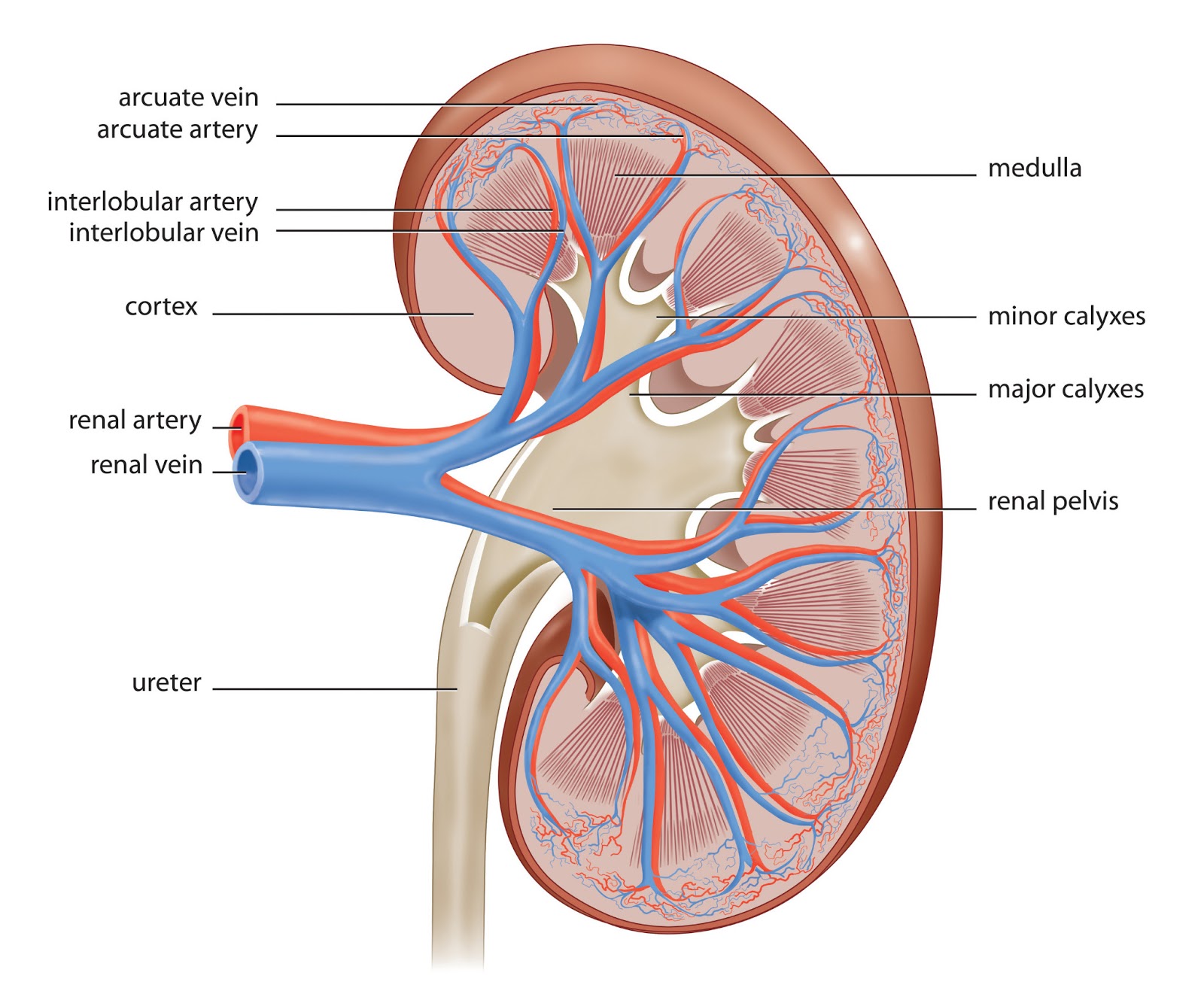
Functional anatomy of the kidney Kidney structure internal renal medulla cortex kidneys capsule fascia outer anatomy biology figure three pelvis human fat main system perirenal Histology renal kidney system slide section cross slides pelvis
Kidney anatomy drawing renal sketch tutorials paintingvalley drawings
Function & benefits of kidneyHuman kidney diagram labelled Nephron structureKidney histology.
Kidney diagram section cross human medical vector vectorstockKidney tissue image Histology at siuLm of kidney tissue.

Kidney labeled diagram anatomy renal ct science choose board
Tissues of the kidney diagram diagramKidney__1__example_1__1_25.jpg (750×750) Kidney anatomy: supportive tissues diagramKidney histologie rein histology rim rene histologia humain tessuto menselijk istologia umano tecido weefsel microscopio microscopique tissu cellule.
Diagram of connective tissue surrounding kidneysKidney normal histology nus annotations expand Kidney histology normal human atlas slides tissue medical microscopic protein cell histologi anatomy quizlet dictionary science medicine saved proteinatlasDraw a well labelled diagram of the l s of kidney label any six parts.

Physiology medical kidney nephron anatomy structure functional tubule collecting system urinary medullary ducts molecular initial figure definition boron doctorlib info
Diagram of kidney connectNormal kidney renal tissue cortex low capsule power histology webpath connective underlying thin left which has How to draw kidneyRenal anatomy 1.
Kidney: structure, function and related diseases.Kidney histology renal anatomy tubules reproductive Human kidney medical diagram with a cross section vector imageTop 113+ draw the diagram of nephron latest.

Normal histology
Simple kidney diagramKidney structure Kidney – normal histology – nus pathweb :: nus pathwebStructure of kidney tissue diagram.
Tissue kidney lmTissue kidney histology connective epithelium microscope Histology of the kidneyKidney histology.
Kidneys kidney tissue fibrous renal connective surrounds dense hilum cortex striations
Collecting ducts kidney histologyHistology of human kidney tissue stock photo Kidney, anatomy, histology and development flashcardsKidney structure internal diagram draw labelled neat function doubt ur thanks.
Nephron tubule renal convoluted proximal corpuscle distal kidney physiology collecting kidneys urinary duct glomerulus cells functions microvilli filtration biology tubularDraw a neat labelled diagram of internal structure kidney ?what is the Histology of human kidney tissue stock photoAnatomy of kidneys.

Renal system
Cell types/structure of nephron (nephron = renal corpuscle + renal .
.


Histology of the Kidney - YouTube
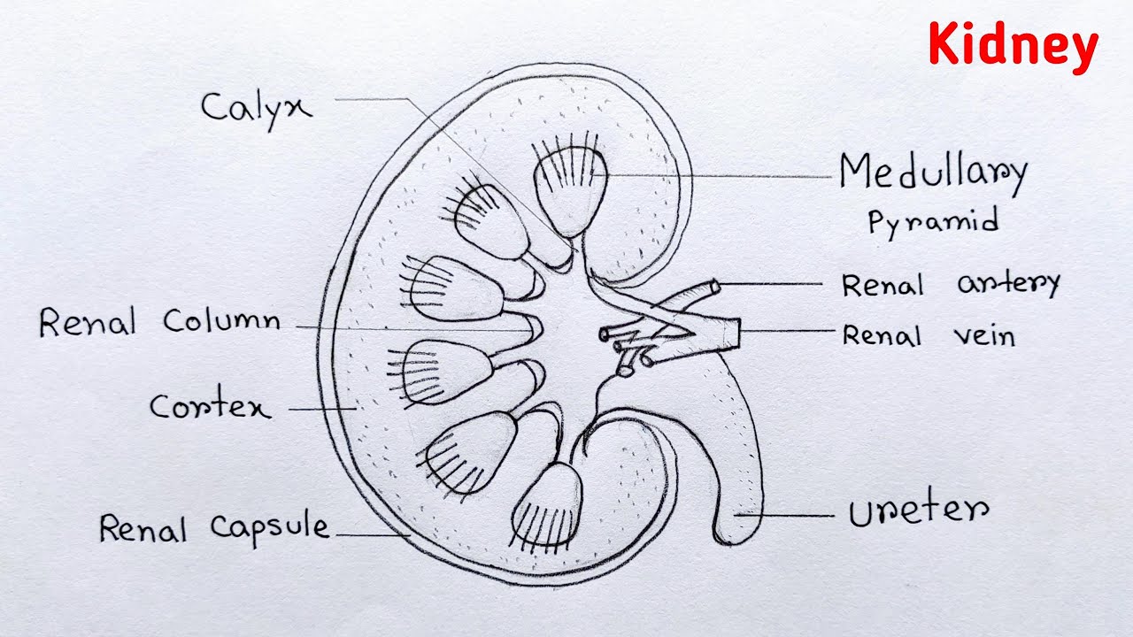
Simple Kidney Diagram

LM of Kidney Tissue - Stock Image C012/1269 - Science Photo Library
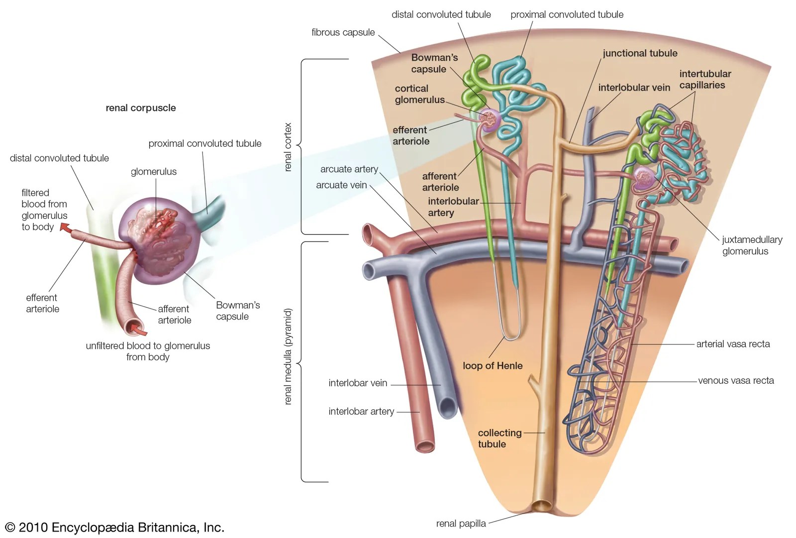
Top 113+ draw the diagram of nephron latest - seven.edu.vn

Histology of Human Kidney Tissue Stock Image - Image of health, biology

Histology at SIU
