Ecg / ekg components Top more than 153 ecg machine drawing latest 1-05. graphic display of electrocardiogram (c)
1-05. GRAPHIC DISPLAY OF ELECTROCARDIOGRAM (C) | Cardiac Rhythm
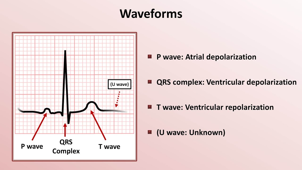
Ecg normal electrocardiography representation wave qrs ekg interval waves complex rhythm electrocardiogram sinus labeled diagram heart cardiac labels ventricular schematic Ecg waveform explained pdf ekg labeled diagrams components waves Basics parts to an electrokardiogram [ekg]
Cardiac ecg interpretation ekg electrocardiogram dysrhythmia impairment heart wave activity electrical nursing graphic learning rhythms patterns course sheet medical rhythm
Mi breviario: ecg o ekgEcg waveform explained pdf: ekg labeled diagrams, components, waves Pin by selina rodriguez on trainingEkg diagram.
Ekg corresponding paramedicine practicingHow to read ecg for nurses Ecg ekg waveform labeledEcg heart rate ekg strip electrocardiogram cardiac howmed interpretation time wave calculation line way nursing mi rhythms tracing information breviario.

Wikipedia electrocardiography wiki
Ecg conduction cardiac system imbalance node electrical cycle impulse sinoatrial electrocardiography ventricles electrolyte atria ekg action potential interpretation changes waveformsRight axis deviation (rad) • litfl • ecg library diagnosis Iot based ecg monitoring with ad8232 ecg sensor and esp32 on ubidotsEcg interpretation: characteristics of the normal ecg (p-wave, qrs.
Ecg components ekg wave monitorElectrocardiogram ecg ekg wave segment waves interpretation rhythm cardiac figure internal graphic display ii Physiology glossary: electrocardiogram (ecg)Axis ecg litfl rad interpretation qrs lad cardiac degrees.
![Basics Parts to an Electrokardiogram [EKG] - YouTube](https://i.ytimg.com/vi/yVtaquWMh2Q/maxresdefault.jpg)
12-lead ekg and the corresponding parts of the... — practicing paramedicine
Ecg atrial pulmonale mitrale enlargement ekg interpretation qrs segment waves contour abnormal interval biphasic amplitude criteria morphology deflection leads notchEkg diagram labeled Nursing school cardiac nurse choose board funny pharmacology ecgEcg normal components simple three grimes david dr shows not.
Ecg waveform explained: ekg labeled diagrams and components — ezmedEkg interpretation (7) diagram 1-05. graphic display of electrocardiogram (d)Pin on mrcp1.

A basic guide to ecg/ekg interpretation
Ecg changes due to electrolyte imbalance (electrolyte disorder) – ecgVarious parts of ecg [diagram] normal ekg diagramEcg leads.
Pin on da ️Ecg ekg interpretation read intervals segments waveforms basics nurses time result tracings Ecg ekg interpretation systematic ecgwaves interpret approach qrs wave learning stEcg waveform explained ekg labeled diagrams and components.

Ekg diagram labeled
[diagram] normal ekg diagramDr david grimes: a simple view of the ecg Ecg parts basics ppt wave powerpoint ventricle downward presentationHow to interpret the ecg / ekg: a systematic approach – ecg learning.
.


ECG / EKG Components

EKG Diagram Labeled
HOW TO READ ECG FOR NURSES - Nursing Manthra

1-05. GRAPHIC DISPLAY OF ELECTROCARDIOGRAM (D) | Cardiac Rhythm

ECG Waveform Explained: EKG Labeled Diagrams and Components — EZmed

ECG Waveform Explained PDF: EKG Labeled Diagrams, Components, Waves

ECG interpretation: Characteristics of the normal ECG (P-wave, QRS
